Cellogram: On-the-Fly Traction Force Microscopy
Abstract
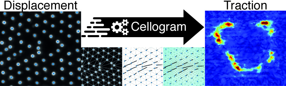
Traction force microscopy (TFM) derives maps of cell-generated forces, typically in the nanonewton range, transmitted to the extracellular environment upon actuation of complex biological processes. In traditional approaches, force rendering requires a terminal, time-consuming step of cell deadhesion to obtain a reference image. A conceptually opposite approach is provided by reference-free methods, opening to the on-the-fly generation of force maps from an ongoing experiment. This requires an image processing algorithm keeping the pace of the biological phenomena under investigation. Here, we introduce an integrated software pipeline rendering force maps from single reference-free TFM images seconds to minutes after their acquisition. The algorithm tackles image processing, reference image estimation, and finite element analysis as a single problem, yielding a robust and fully automatic solution. The method’s capabilities are demonstrated in two applications. First, the mechanical annihilation of cancer cells is monitored as a function of rising environmental temperature, setting a population threshold at 45 °C. Second, the fast temporal correlation of forces produced across individual cells is used to map physically connected adhesion points, yielding typical lengths that vary as a function of the cell cycle phase.
Citation
@article{Lendenmann:2019:COT,
    author = {Lendenmann, Tobias and Schneider, Teseo and Dumas, Jérémie and Tarini, Marco and Giampietro, Costanza and Bajpai, Apratim and Chen, Weiqiang and Gerber, Julia and Poulikakos, Dimos and Ferrari, Aldo and Panozzo, Daniele},
    doi = {10.1021/acs.nanolett.9b01505},
    eprint = {https://doi.org/10.1021/acs.nanolett.9b01505},
    journal = {Nano Letters},
    note = {PMID: 31538794},
    title = {Cellogram: On-The-Fly Traction Force Microscopy},
    url = {https://doi.org/10.1021/acs.nanolett.9b01505},
    year = {2019}
}
Acknowledgments
We thank Francesca Michela Pramotton, Raoul Hopf, and Magaly Reyes for input during analysis and Sally Reynolds for her artistic interpretation of Cellogram leading to the app icon. We acknowledge the Computational Image Analysis in Cell and Developmental Biology in Woods Hole for providing T.L. with C code for the detection of particles.
T.L. was supported by funding from the ETH-grant ETH-12 15–1. This work was partially supported by the NSF CAREER award 1652515, the NSF grant 1835712, the SNSF grant P2TIP2_175859, a gift from Adobe Research, the American Heart Association Scientist Development Grant (16SDG31020038).